Pielea spune dintr-o privire povestea fiecăruia dintre noi, cu informații despre afecțiunile, nutriția, vârsta și obiceiurile noastre. Cum o impresie bună se face cu o piele sănătoasă și bine îngrijită, vă asigurăm că în SKINMED® găsiți profesioniști, tehnologie și experiență pentru ca aspectul dumneavoastră să fie impecabil.
În SKINMED®, dorim să fiți nu doar cea mai sănătoasă și frumoasă versiune a dumneavoastră, ci și cea mai mulțumită.
Servicii
Soluții complete pentru toate zonele corpului
Echipa SKINMED®


Centre de Excelență
Blog
HArmonyCa, acidul hialuronic cu efect de lifting instant & susţinut!
HArmonyCa este un produs injectabil hibrid cu efect dublu. Noul filler conţine două ingrediente active, hidroxiapatita de calciu (CaHa) şi acidul...
Citește mai mult

Elimină bărbia dublă (gușa) fără bisturiu!
Bărbia dublă, popular cunoscută drept gușă, înrăutățește vizibil aspectul unui chip. Indiferent de cauze, fie că vorbim despre înaintarea în...


HArmonyCa, acidul hialuronic cu efect de lifting instant & susţinut!
HArmonyCa este un produs injectabil hibrid cu efect dublu. Noul filler conţine două ingrediente active, hidroxiapatita de calciu (CaHa) şi acidul...
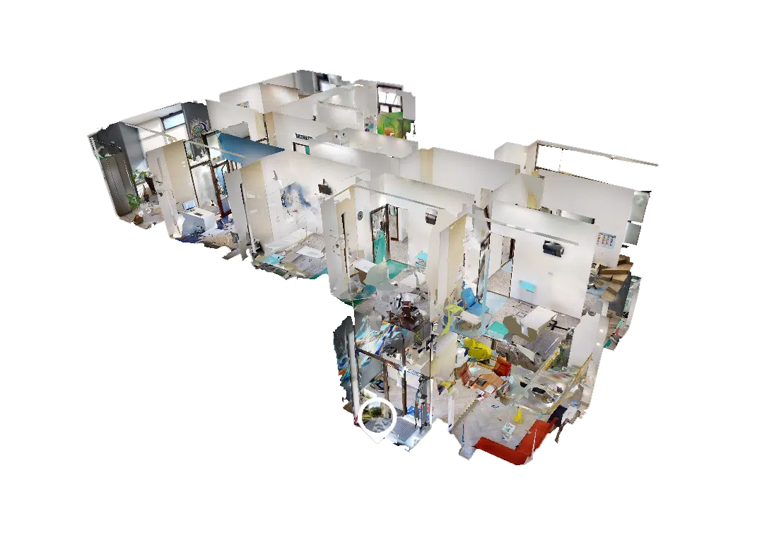
Vizitează SKINMED® în 3D!
Anul 2023
26174
de tratamente
3472
de pacienți noi
2351
de tratamente epilare laser
1913
de tratamente injectabile

Parteneri